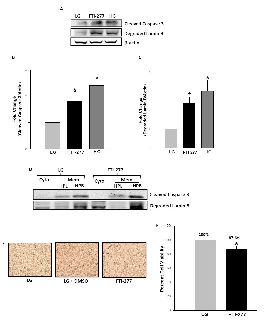
Fig. 3. FTI-277 induces caspase-3 activation, lamin degradation and loss in cell viability in INS-1 832 cells. Panel A: INS-1 832/13 cells were incubated in a medium containing low glucose (2.5 mM; LG) in the absence and presence of FTI-277 (10 µM) for 24 hrs. Caspase 3 activation and lamin B degradation for each condition was determined by Western blotting (Panel A). Equal protein loading was ensured by re-probing the membrane with anti β-actin. Intensity of protein bands was quantified by densitometry. Data represent mean ± SEM from three independent experiments, and are expressed as fold change in caspase 3 activation (Panel B) and lamin B degradation (Panel C). *P<0.05 vs. LG. Panel D: Lysates from INS-1 832/13 cells treated with low glucose (2.5 mM) in the absence and presence of FTI-277 (10 µM) were subjected to a single-step centrifugation to separate the supernatant cytosolic (Cyto) and the membrane-pellet (Mem) fractions followed by hydrophobic (HPB) and hydrophilic (HPL) phase partitioning with Triton X-114 detergent [23,24]. The lysate proteins were resolved by SDS-PAGE and transferred to a nitrocellulose membrane. The membrane was probed for cleaved caspase 3 and lamin B. A representative blot from two independent experiments is shown here. Panel E: INS-1 832/13 cells were treated with media containing low glucose (2.5 mM; LG) alone and in the presence of diluent (DMSO), FTI- 277 (10 µM) for 24 hrs. Changes in cell morphology were visualized by light microscopy. Panel F: INS-1 832/13 cells were treated with low glucose (2.5 mM; LG) in the absence and presence of FTI-277 (10 µM). After 24 hrs. cell viability was assessed by the MTT assay. Data represent mean ± SEM and are expressed as percent cell viability. *P<0.05 vs. LG.